Research Directions
The Vahabikashi Lab pursues basic science and translational research at the intersection of multiscale mechanobiology and soft bioelectronics. Our projects span from fundamental investigations of mechanobiology across nuclear, cellular, and tissue scales in health and disease, to the engineering of next-generation microphysiological systems, and implantable devices for mechanobiology studies and regenerative engineering.
Current work in the lab focuses on four project areas:
Mechanobiology of Ocular Hypertension and Glaucoma: We investigate how altered mechanobiology of Schlemm's canal inner wall endothelium and the juxtacanalicular tissue aspect of trabecular meshwork drives elevated intraocular pressure in glaucoma, and we develop targeted therapies to restore normal outflow resistance.
Nuclear Mechanobiology and Mechanotransduction: We study how nuclear lamin intermediate filaments contribute to cell mechanics, mechanotransduction, and migration, both as a fundamental biological question and in the context of cancer and laminopathies.
Soft 3D Bioelectronic Interfaces for Organoid and Tissue-Based Microphysiological Systems: We integrate flexible, multimodal bioelectronic interfaces within microfluidic devices to create organ- and organoid-on-chip models of ocular and neuromuscular systems from iPSC-derived tissues, which enable simultaneous electrophysiology, contractility, and force measurements alongside optical monitoring via high-resolution microscopy.
Implantable and Wearable Bioelectronics for Disease Studies and Tissue Regeneration: We engineer flexible and stretchable bioelectronic devices with wireless power and data transfer, electrical stimulation, and multimodal sensing, and integrate them with stem cell–derived tissues for cardiac and neural regeneration. Current projects include cardiac patches for myocardial repair, smart conduits for guided retinal ganglion cell growth and optic nerve regeneration, and implantable/wearable ocular telemetry devices for wireless monitoring of intraocular pressure, intracranial pressure, and electrophysiological activity in animal models of ocular neurodegenerative diseases such as glaucoma.
Click on “Read More” to learn more about the projects
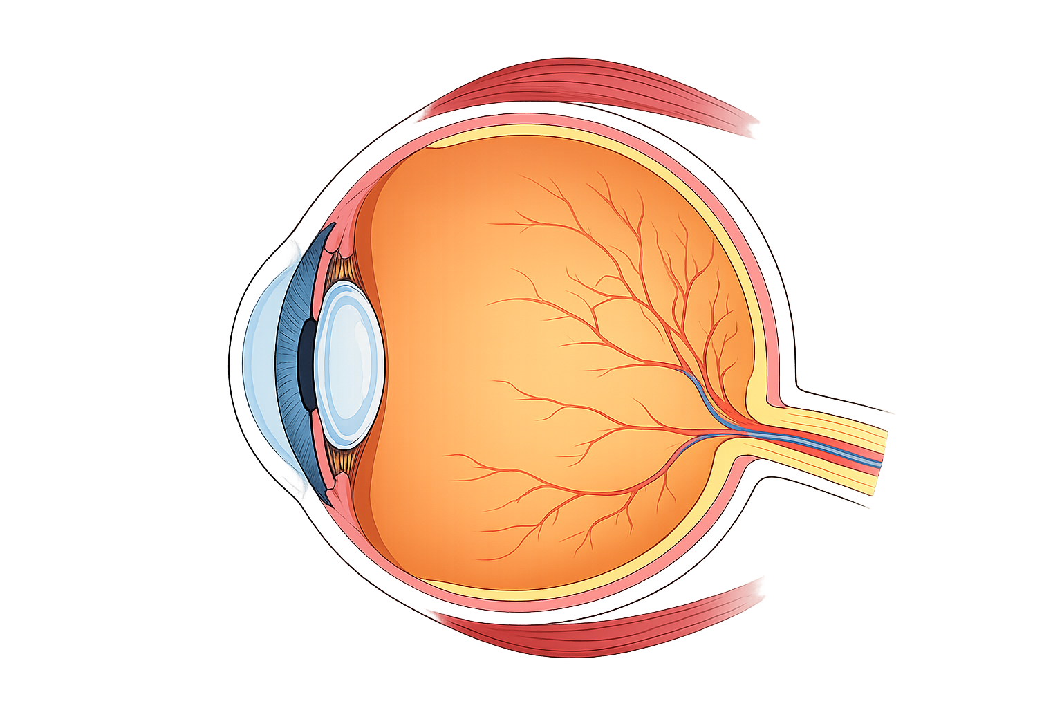
Glaucoma Mechanobiology
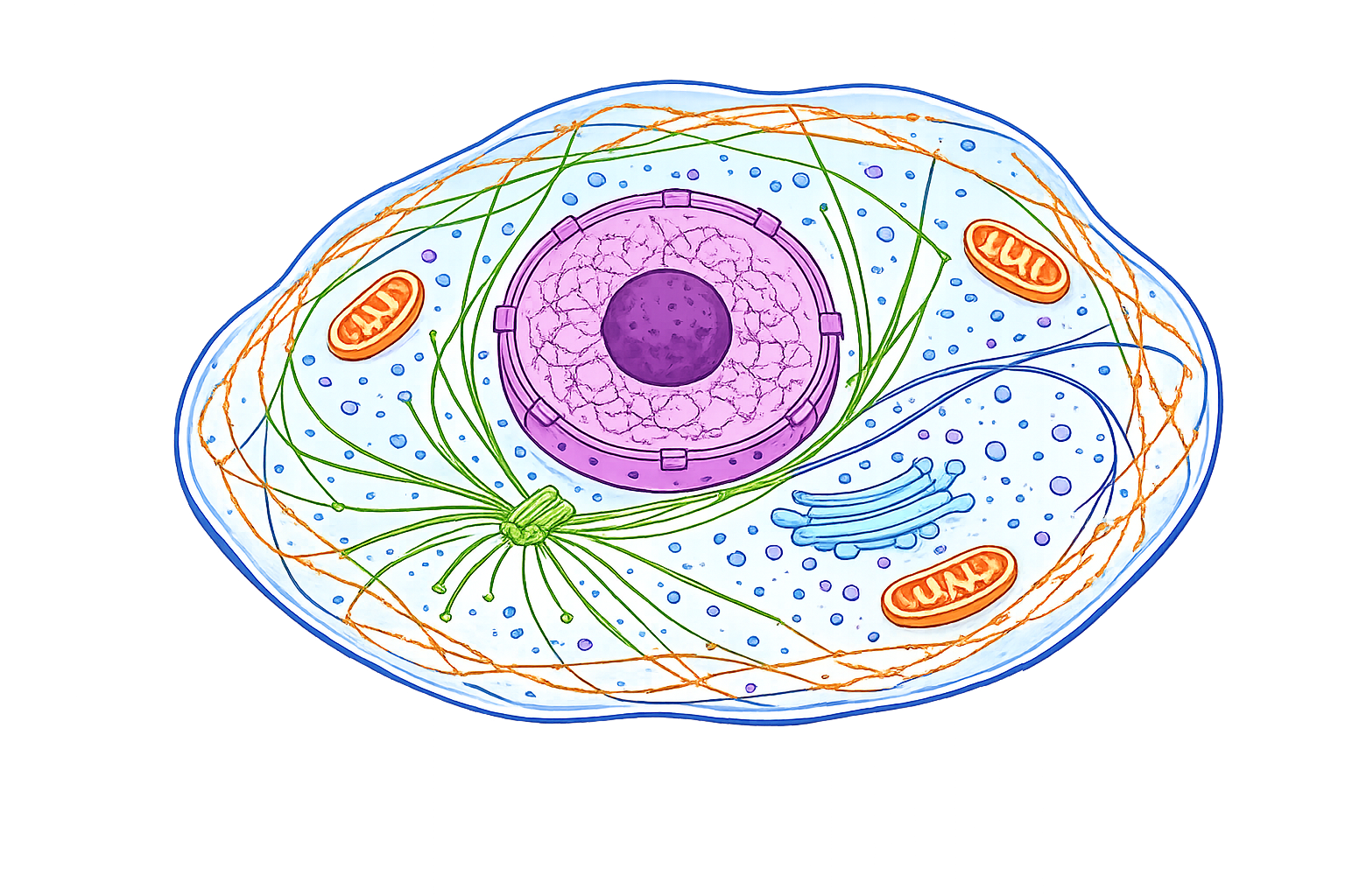
Nuclear Mechanobiology
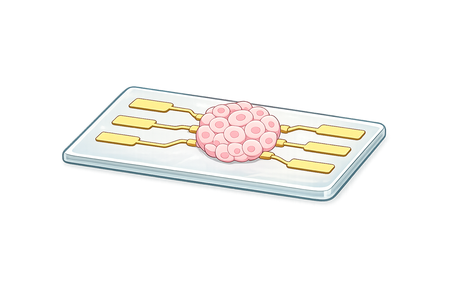
Organ-on-Chip Bioelectronics