
Mechanobiology of Ocular Hypertension and Glaucoma
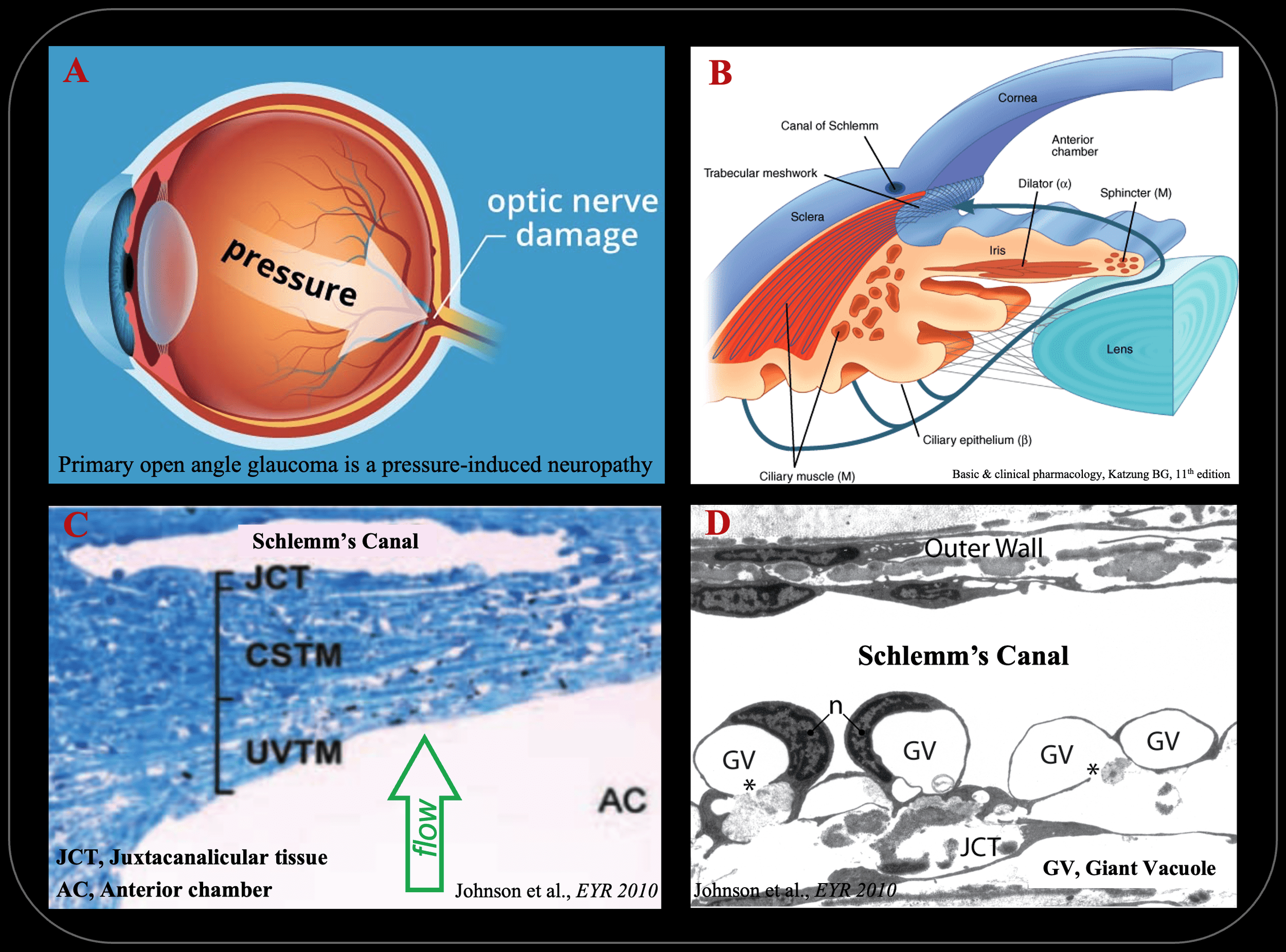
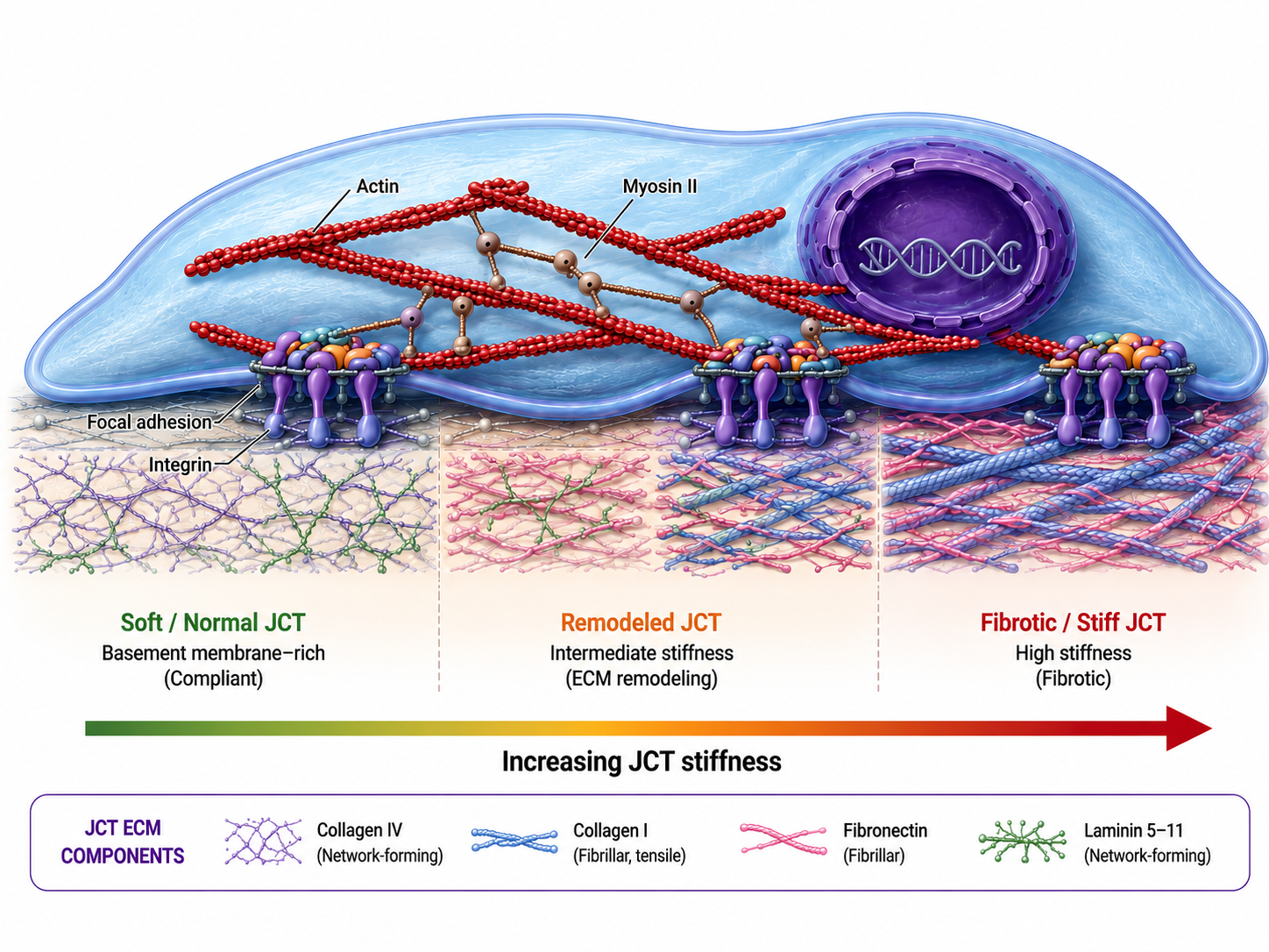
Glaucoma is a family of neuropathies and the leading cause of irreversible blindness in the world. Primary open-angle glaucoma (POAG) is the largest subset of the disease, and its elevated intraocular pressure characteristic is attributable to increased resistance to the aqueous humor outflow from the eye. However, the underlying mechanisms of this increased flow resistance and its primary location have eluded investigators for over 140 years. We have previously shown that increased stiffness of Schlemm's canal (SC) endothelium and its underlying substrate, the juxtacanalicular tissue (JCT) of the trabecular meshwork (TM), is central to the elevated outflow resistance and intraocular pressure in human glaucomatous eyes. Nonetheless, the underlying processes driving this etiology remain poorly understood.
We aim to investigate the mechanisms responsible for altered mechanobiology of SC endothelium and the TM, and to leverage this understanding to develop new therapeutics and regenerative approaches for the disease. A representative example of these efforts is our demonstration that targeted delivery of nanoparticles that soften SC endothelial cells significantly reduces intraocular pressure in mouse models, suggesting that targeting SC and TM mechanobiology could be a promising avenue for developing new glaucoma therapies.
Graduate students and RAs working on the project:
Disease mechanism - Daniel Labbe, Brooke Zhou, Aadhira Nair
Organ-on-a-chip model systems - Keerthi Atluri